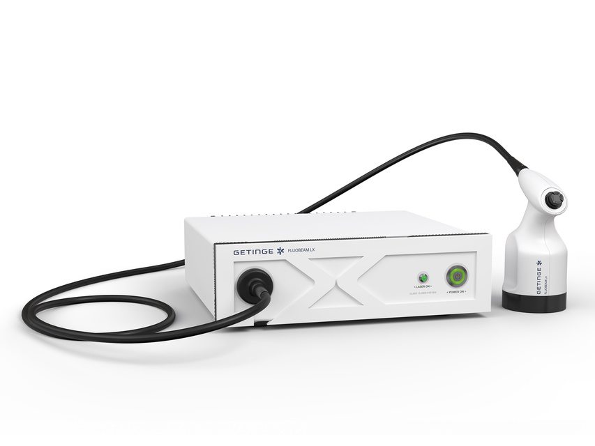
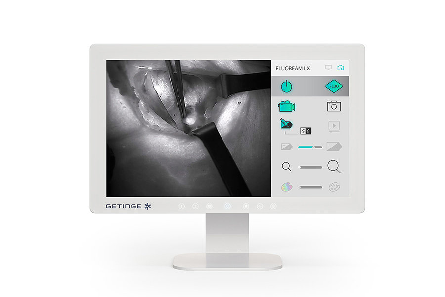
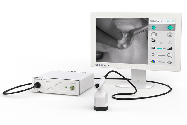
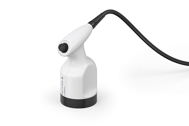
FLUOBEAM LX
EMIG GmbH - Official distributor of medical equipment in the following regions of Europe and beyond:
- Meyer & Haake – Ukraine, Austria, Germany (Bavaria); RiwoSpine – Ukraine, Moldova, Estonia;
- Inomed – Ukraine, Moldova, Estonia; Fluoptics – Ukraine.
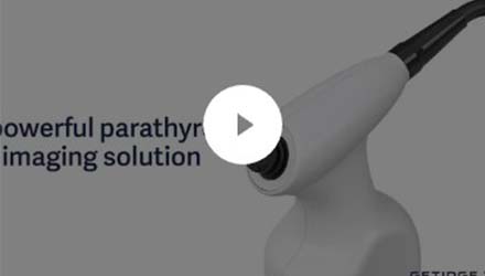
A dedicated camera for thyroid and parathyroid surgery
FLUOBEAM LX is an imaging device exclusively dedicated to thyroid and parathyroid surgery, offering surgeons optimal comfort during use throughout the procedure. Its ease of use and ability to analyze images make it a major asset for surgeons.
A powerful parathyroid imaging solution
FLUOBEAM LX combines autofluorescence and fluorescence perfusion imaging to provide surgeons with enhanced visualization during surgery.
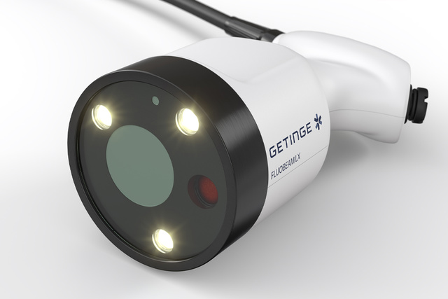
Fluorescence imaging device
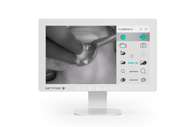
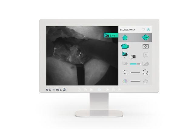
FLUOBEAM LX is a fluorescence imaging device dedicated to thyroid and parathyroid surgery. It provides both autofluorescence and Indocyanine green ICG fluorescence imaging.
Autofluorescence
Highly sensitive, FLUOBEAM LX allows to early detect parathyroid glands by autofluorescence with an optimized real-time display, a high depth of field and a compatibility with ambient operating room lights.
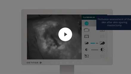
Perfusion assessment
Parathyroid glands perfusion assessment at the end of surgery, provides a posteriori information on the quality of surgery and also informs on possible postoperative complications.
Real-time imaging
The FLUOBEAM LX allows for the detection and imaging of the parathyroid glands in real time (25 frames/second) and in ambient light thanks to an optimized optical filtering. With the surgical lights on, autofluorescence can still be used by turning the lights away from the incision.[1] This allows an easier integration of autofluorescence in the surgical workflow.
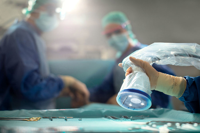
A clear image from 8 to 13 cm
To ensure a clear image, the camera operates with a very large depth of field not forcing the surgeon to adjust the focus. The focusing distance and large depth of field correspond to a natural holding of the camera above the incision and maintains the necessary sharpness of the area of interest to locate weak signals of the parathyroid glands.
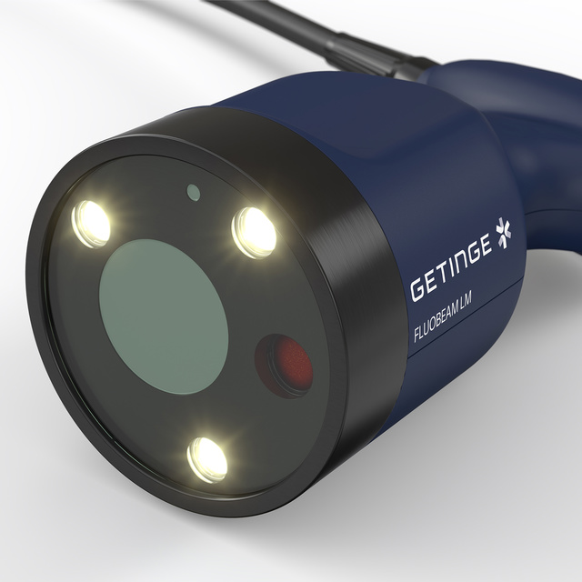
Class 1 laser
Most existing fluorescence imaging systems use Class 3R laser illumination which can be dangerous under certain operating conditions. Our class 1 laser ensures total safety for the eyes of the users. This choice of optical safety was made without compromising the unmatched sensitivity of the system.

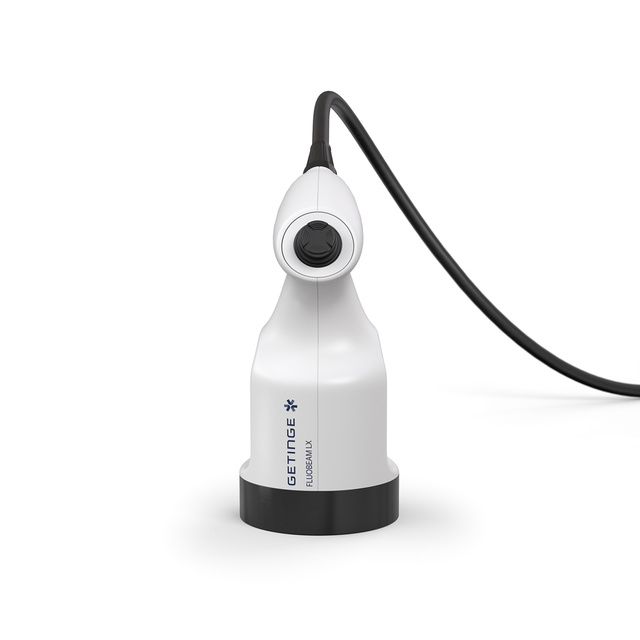
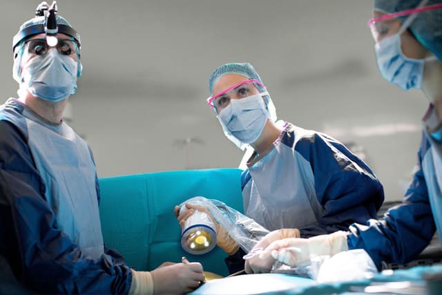
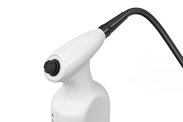
Joystick control
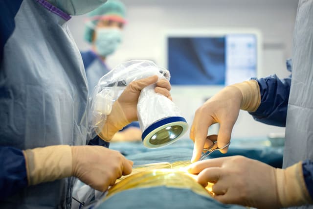
Designed to be easily held and manipulated in the surgical field, FLUOBEAM LX offers optimized ergonomics with a joystick that simplifies the navigation and the selection of the software functionalities, directly by the surgeon.
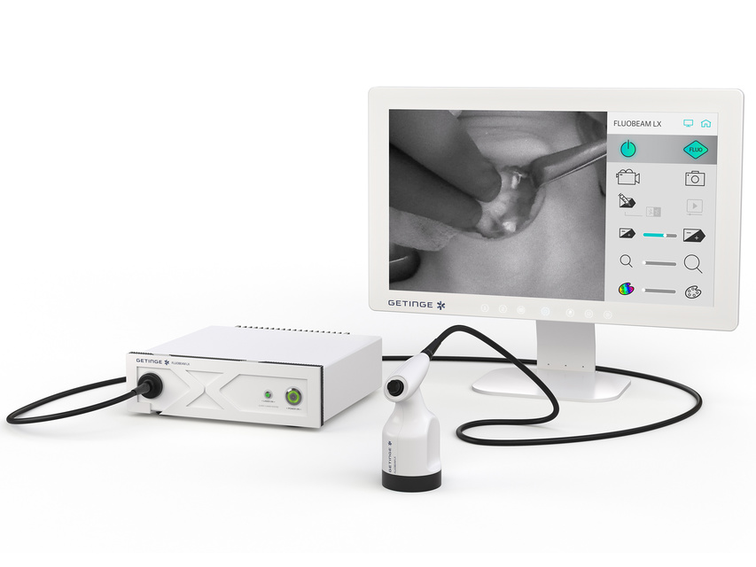
Unrivaled images
With a high frame rate for real-time display (25 frames/s) in autofluorescence and a high depth of field (> 5cm), with FLUOSOFT LX imaging software, surgeons can work in optimized conditions with an easy interpretation of images and manipulation of the device.
Fluorescence imaging brings benefits in thyroid surgery
Reduction of transient hypocalcemia
Several clinical teams using FLUOBEAM LX have demonstrated that the use of autofluorescence as a tracking tool during thyroid surgery leads to better preservation of the parathyroid glands and their functions. This leads to a significant reduction in the rate of transient post-operative hypocalcemia.
A clear image from 8 to 13 cm
On average, pathologists find that a parathyroid gland is inadvertently removed in 10% to 25% of thyroidectomies.
With the use of autofluorescence imaging, the parathyroid glands are early detected and can be preserved in most cases.
Class 1 laser
Autofluorescence can be coupled with the use of fluorescence imaging by injection of indocyanine green. This allows the identification of the vessels that supply the parathyroid glands and thus avoids any risk of devascularization.
In cases where the parathyroid glands could not be preserved, their identification by autofluorescence makes it possible to locate them in the removed specimen and to proceed to their reimplantation.
Intraoperative fluorescence imaging, a precise and efficient method
Parathyroid glands identification can be challenging even for experienced surgeons due to their tiny size (a few mm) and that they can be ectopic our buried.
The unexpected excision of healthy parathyroid glands is a current complication of thyroidectomies. This can lead to hypoparathyroidism, most of the time transient and might result in disruptions of calcium metabolism and notably hypocalcemia. It is therefore critical to properly identify parathyroid glands perfusion assessment during surgery.
Thyroid surgery
Parathyroid gland visualization
Parathyroid glands naturally emit fluorescence in the near infrared without any dye injection. This is called autofluorescence.
FLUOBEAM LX helps the surgeon visualize in real-time parathyroid glands and preserve them during surgery.
Identification of vessels that perfuse parathyroid glands
By easily combining autofluorescence and indocyanine green angiography during the procedure, FLUOBEAM LX allows a clear identification of vessels that perfuse parathyroid glands and assists with their preservation during thyroid dissection.
Checking the parathyroid gland vascularization
It is commonly known that complications such as transient hypocalcemia are linked to the unexpected excision of parathyroid glands or the alteration of the vascularization of these glands during thyroid surgery.
After intravenous injection of indocyanine green, FLUOBEAM LX enables surgeons to clearly visualize the vascularization and perfusion of the parathyroid glands, and therefore to assess their viability at the end of the surgery.


An innovative camera for thyroid and parathyroid surgery
- Use of Indocyanine Green Fluorescence During Total Thyroidectomy to Identify Parathyroid Glands and Prevent Hypoparathyroidism. Surg Technol Int. 2023 Dec 29;43 Zhang D, Sun H, Frattini F, Kim HY, Wu CW, Donatini G, Cestari A, Bertoli S, Barbieri D, Bussi M, Dionigi G.
- Intraoperative near‐infrared imaging of parathyroid glands: A comparison of first‐ and second‐generation technologies. Akbulut S. et al. J Surg Oncol. 2020;1–6.
- Impact of autofluorescence-based identification of parathyroids during total thyroidectomy on postoperative hypocalcemia: a before and after controlled study, Benmiloud et al., Surgery 163 (2018) 23–30
- Impact of near-infrared fluorescence imaging plus indocyanine green fluorescence on postoperative hypoparathyroidism rates after total thyroidectomy and central neck lymph node dissection, Di Lorenzo et al., BJS, 2024, znae022
- The use of near‑infrared autofluorescence during total laryngectomy with hemi‑ or total thyroidectomy. Diego Barbieri, Michela Nicole Melegatti, Alessandro Vinciguerra, Pietro Indelicato, Leone Giordano, Stefano Bondi, Matteo Biafora, Matteo Trimarchi, Mario Bussi. European Archives of Oto-Rhino-Laryngology. 2022 August 03
- Association of Autofluorescence-Based Detection of the Parathyroid Glands During Total Thyroidectomy With Postoperative Hypocalcemia Risk Results of the PARAFLUO Multicenter Randomized Clinical Trial
Related products