EMIG GmbH - Official distributor of medical equipment in the following regions of Europe and beyond:
- Meyer & Haake – Ukraine, Austria, Germany (Bavaria); RiwoSpine – Ukraine, Moldova, Estonia;
- Inomed – Ukraine, Moldova, Estonia; Fluoptics – Ukraine.
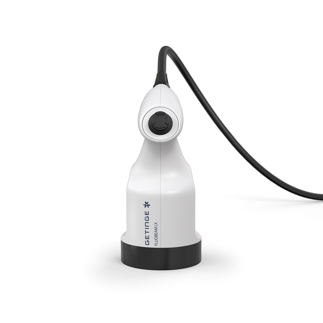
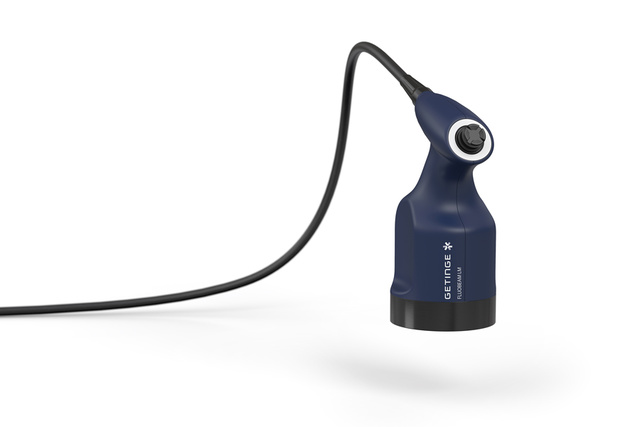
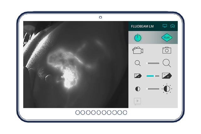
FLUOBEAM LM

Advanced technology to assess perfusion
Building on a decade of experience in fluorescence imaging, FLUOBEAM LM is an integrated fluorescence imaging solution for perfusion assessment.
It provides surgeons with a real-time image of the fluorescence in the operative field.
Advanced technology to assess perfusion
FLUOBEAM LM is dedicated to evaluating perfusion and lymphatic system. When used with Indocyanine Green (ICG), FLUOBEAM LM is an integrated fluorescence imaging solution that provides surgeons with a real-time image of tissue perfusion in fluorescence in the surgical field.
Thousands of procedures already performed.
- Plastic and reconstructive surgery
- Parathyroid gland identification using autofluorescence and perfusion assessment
- Lymphedema and wound treatment
- Partial hepatectomy and liver transplantation
- Sentinel lymph node biopsy for breast cancer and melanoma patients
Excellence at your fingertips
FLUOBEAM LM is an integrated fluorescence imaging solution that provides the surgeon, during surgery, with information invisible to the naked eye such as tissue perfusion, lymphatic drainage status, or lymph node location. It helps the surgeon in his decision-making process in order to reduce the rate of complications.[1],[2],[3] Its ease of use and image processing functions makes it a major asset for surgeons.
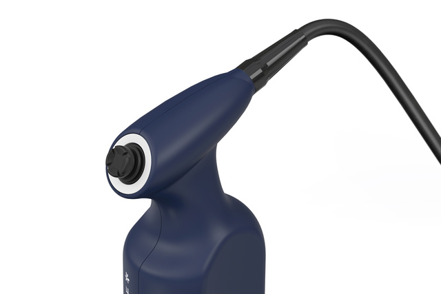
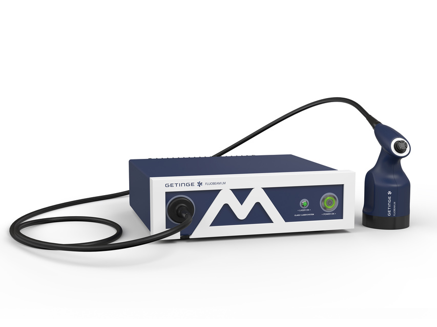
Ergonomics: a certain taste of freedom
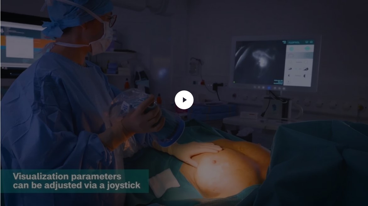
FLUOBEAM LM has been designed to be integrated in the surgical workflow. It offers the surgeon total autonomy during surgery, allowing him to control all acquisition and visualization parameters from the sterile area.
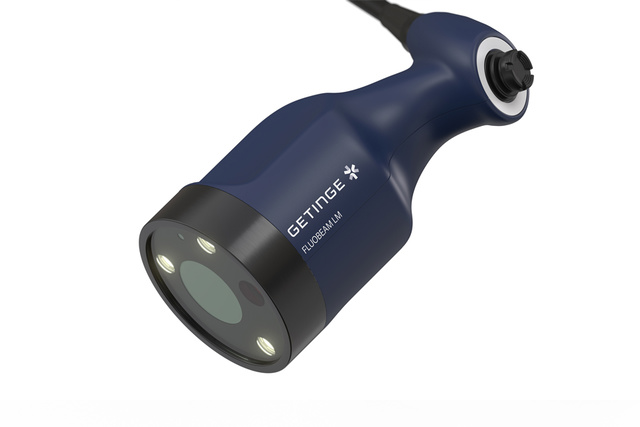
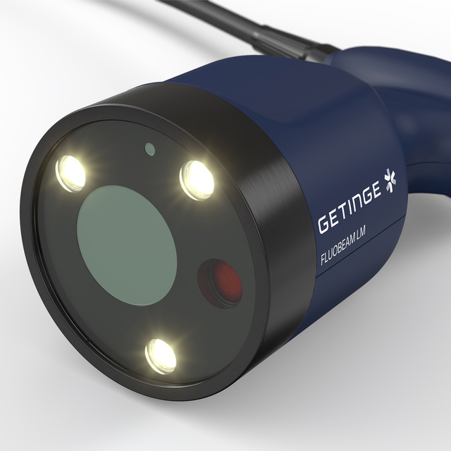
Safety: you won’t believe your eyes
FLUOBEAM LM is an optically safe and ergonomic system. The camera combines safety and performance. Its optical head with a sensitive near-infrared camera excites the fluorescent agent with a class 1 laser, which is harmless to the eye even in direct vision.
Ergonomic and intuitive
The FLUOBEAM LM technology was developed to provide surgeons with a comfortable ergonomic camera and intuitive software during surgery.
Its joystick allows a fluid navigation in the software functionalities and offers the surgeon a total autonomy when using the device.

High performance and safety
FLUOBEAM LM has been designed to offer a high level of technical performance while ensuring surgeon and patient safety.
The power of its Class 1 laser and the quality of its optical filtering allow the surgeon to operate up to a working distance of 12 cm.
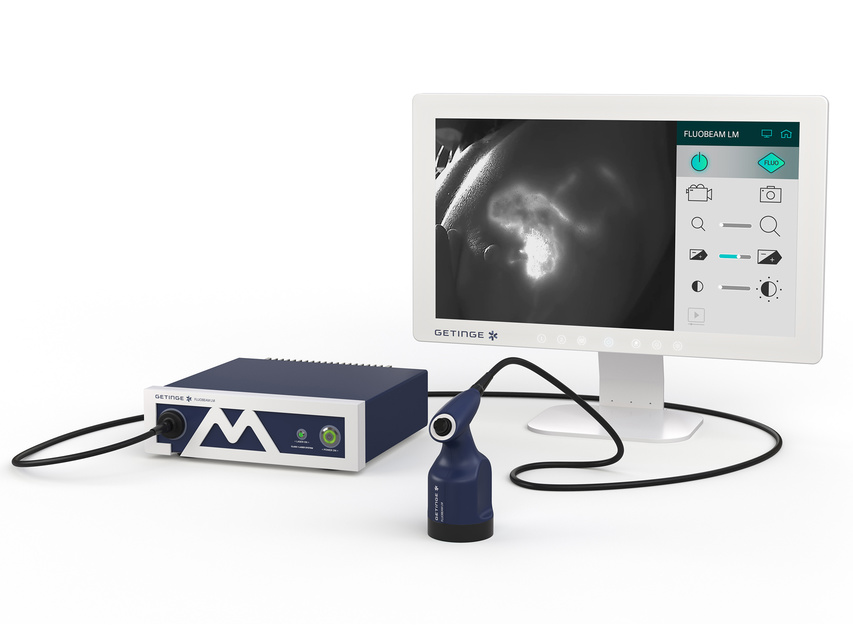
Automatic acquisition mode
FLUOBEAM LM offers more than just images. Its integrated multi-indication software launches with a single click and automatically accesses dynamic information that is difficult to integrate with the naked eye.
The camera allows the surgeon to have direct and autonomous access to an analysis of the perfusion dynamics in the field, the location of the perforator vessels and the extent of their perfusion zone.
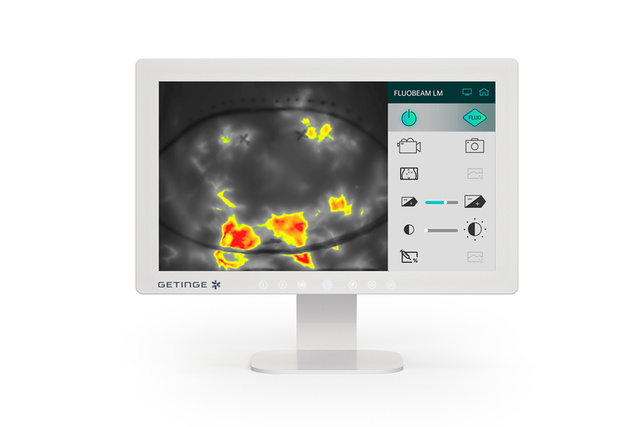
The Most Powerful Solution for Parathyroid Gland Visualization
Operates under standard operating room lighting
Precise identification of parathyroid glands using autofluorescence
Optimized real-time display (high frame rate)
Perfusion assessment of parathyroid glands through indocyanine green injection
Intuitive control (easy one-handed operation)
Aids in clinical decision-making
Combined use of autofluorescence detection and perfusion assessment
Autofluorescence detection reduces the incidence of hypocalcemia after total thyroidectomy
-
Fluorescence Imaging During Surgery: A Precise and Effective Method
Identifying parathyroid glands can be challenging even for experienced surgeons due to their small size (only a few millimeters) and their frequent location within fatty tissue or in atypical positions.
Accidental removal of healthy parathyroid glands is a common complication of thyroidectomy. This may lead to hypoparathyroidism, which is often temporary but can disrupt calcium metabolism and, in most cases, cause hypocalcemia.
Therefore, accurately identifying the parathyroid glands during surgery is critically important.
-
Real-Time Parathyroid Gland Identification
After several years of development in collaboration with international teams of clinical specialists, the FLUOBEAM®LX system has been designed to seamlessly integrate into the operating room environment. FLUOBEAM®LX is a comprehensive fluorescence imaging solution that provides the surgeon with real-time fluorescent images of the surgical field.
The system's ease of use and image analysis capabilities make it a crucial tool for surgeons. There is no assistance available for parathyroid gland localization, and even for experienced surgeons, using FLUOBEAM®LX provides significant help.
-
Fluorescence Imaging During Surgery: A Precise and Effective Method
Identifying parathyroid glands can be a challenge even for experienced surgeons due to their small size (only a few millimeters) and their often being located within fatty tissue or in atypical positions.
Accidental removal of healthy parathyroid glands is a common complication of thyroidectomy. This can lead to hypoparathyroidism, which is usually temporary but can disrupt calcium metabolism and, in most cases, cause hypocalcemia.
Therefore, it is critically important to accurately locate the parathyroid glands during surgery.
-
Identification of Parathyroid Glands
The parathyroid glands are unique organs capable of fluorescing in the near-infrared range without the need for any dyes. This phenomenon is known as autofluorescence. FLUOBEAM®LX allows the surgeon to locate the parathyroid glands in real-time and preserve them during surgery.
FLUOBEAM®LX also enables surgeons to visualize parathyroid adenomas using autofluorescence. This identification guides the surgeon and facilitates the resection process.
-
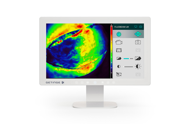
Verification of Parathyroid Gland Vascularization
It is well known that complications, such as transient hypocalcemia, are associated with the accidental removal of parathyroid glands or disruption of their vascularization during thyroid surgery.
After intravenous injection of indocyanine green, FLUOBEAM® allows surgeons to see a clear image of the vascularization of the parathyroid glands, thereby assessing their viability during the surgery.
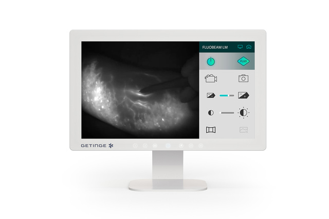
Plastic and Reconstructive surgery
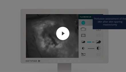
FLUOBEAM® LM provides surgeons with accurate information to help locate perforator vessels during free flap reconstruction procedures and to assess tissue perfusion during reconstructive surgeries and after skin-sparing mastectomies.
Axillary sentinel lymph node biopsy
FLUOBEAM® LM provides surgeons with accurate information to easily detect and remove sentinel lymph nodes in patients with breast cancer.
Visualization of lymphatic drainage
FLUOBEAM® LM provides surgeons with real-time visualization of superficial lymphatic drainage to assess and analyze its efficiency and/or dysfunction in patients with lymphedema.
An indication-oriented software
Fluorescence imaging brings benefits in
Plastic and Reconstructive surgery
- Intraoperative identification of perforator vessels and the perforator angiosome
- Real-time intraoperative tissue perfusion assessment
- More precise flap design according to the perfused areas
- Relative quantification tool (additional information to improve the specificity of the method)
- Early identification of complications
- Postoperative monitoring
Axillary sentinel lymph node biopsy
- High detection rate (similar to radioisotope)
- Direct visualization (lymphatic drainage and sentinel lymph node)
- Simple patient care pathway (one injection only on the day of surgery)
- No need for radioactive marker injection
- Risk reduction (low allergy rate compared to blue dye)
Lymphatic drainage visualization
- Early identification and localization of lymphatic drainage deficiencies to establish appropriate treatment (surgical or not)
- Accurate visualization of functional lymphatic vessels before lymphaticovenous anastomosis (LVA)
- Accurate location of lymph nodes during lymph node transfer surgery
- Post-treatment monitoring
- Reverse mapping capability
Fluorescence imaging in plastic and reconstructive surgery
Plastic and reconstructive surgeries require precise assessment of tissue perfusion quality to prevent partial necrosis. Indocyanine Green (ICG) fluorescence angiography offers reliable verification of tissue perfusion and identification of at-risk areas. FLUOBEAM LM facilitates real-time visualization of ICG flow, distribution, and accumulation on a screen during surgery, aiding in visualizing blood flow.
Breast reconstruction
FLUOBEAM LM allows the surgeon to evaluate in real time the quality of tissue perfusion (flaps or skin after mastectomy with preservation of the skin flap) and to adapt the surgical procedure to minimize the risks of complications.
During a reconstruction procedure, with the use of an autologous flap (free or pedicled), it is essential to ensure good tissue perfusion to avoid postoperative complications, such as partial or fat necrosis.
Fluorescence imaging in lymphatic surgery
In lymphatic surgery, it is essential to precisely locate functional lymphatic vessels. During consultation and surgery FLUOBEAM LM is used to image in real time the quality of the lymphatic network.
Observation of lymphatic vessels
In medical consultation, FLUOBEAM LM can be used for real-time imaging of functional superficial lymphatic vessels. It helps in the early diagnosis of lymphedema, in the choice of treatment (surgical or medical), and in the postoperative follow-up.
In surgery, FLUOBEAM LM can be used to identify before and during surgery the functional lymphatic vessels that will be used for a lymphaticovenous anastomosis (LVA). In addition, FLUOBEAM LM can be used in “reverse mapping” to map and preserve lymphatic drainage during lymph node surgery and can also be used for postoperative follow-up.
Sentinel lymph node biopsy
The sentinel lymph node is the first lymph node where cancer cells are most likely to migrate from the primary tumor. The sentinel node technique involves detecting this node, removing it and having it analyzed.
Fluorescence imaging and Indocyanine Green (ICG) have been recently introduced in the clinic as an alternative for surgical guidance. FLUOBEAM LM allows visualization of superficial lymphatic drainage and detection of the sentinel lymph node after incision, for different tumor types.


Advanced technology to access perfusion
- Prospective Clinical Trial for Predicting Mastectomy Skin Flap Necrosis with Indocyanine Green Angiography in Implant-Based Prepectoral Breast Reconstruction. Aesthetic Plast Surg. 2024 May 13. Jaewoo Kim, Man Wong Han, Ki Yong Hong.
- Mapping of the superficial lymphatic network of the breast using indocyanine green injection: an anatomical study. Research Square 2022. Yannis CLAUDIC, Anne PERRUISSEAU-CARRIER, Charlie DEMARTELEIRE, Weiguo HU and Romuald SEIZEUR.
- Indocyanine green for sentinel lymph node detection in early breast cancer: Prospective evaluation of detection rate and toxicity-The FLUOBREAST trial Breast J. 2020 Oct 22. C. Ngô, S. Sharifzadehgan, C. Lecurieux-Lafayette, H. Belhouari, D. Rousseau, H. Bonsang-Kitzis, L. Crouillebois, V. Balaya, S. Oudard, F. Lécuru, R-T. Elaidi.
- Preventing Soft Tissue Complications in Secondary Aesthetic Breast Surgery Using Indocyanin Green Angiography. Salgarello M. et al. Aesthetic Surgery Journal 2023, Vol 43(6) 665–672
- Prospective Clinical Trial for Predicting Mastectomy Skin Flap Necrosis with Indocyanine Green Angiography in Implant-Based Prepectoral Breast Reconstruction. Kim J. 2024 Aesth Plast Surg